
Digital Mammography for Breast Cancer Detection
Digital mammography is one of the most established imaging methods for detecting breast cancer at an early stage. It uses low-dose X-rays to produce high-resolution images of the breast, allowing radiologists to identify abnormalities such as masses, architectural distortions, and suspicious calcifications that may be associated with malignancy.
Unlike traditional film-screen mammography, digital mammography captures breast images electronically, allowing them to be processed, enhanced, stored, and reviewed using advanced digital systems. This improves image accessibility and supports detailed interpretation by breast imaging specialists.
How Does Digital Mammography Help Detect Breast Cancer?
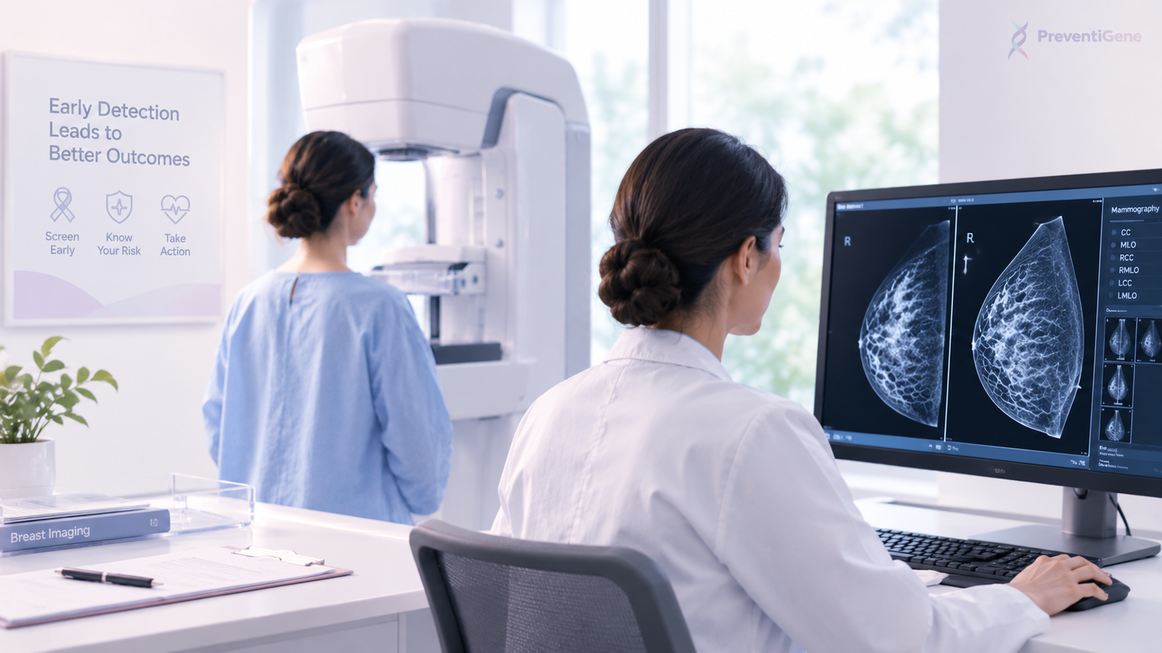
During a mammographic examination, the breast is gently compressed between two plates. X-ray images are then acquired from different standard views. Compression helps reduce tissue overlap and improves image quality while keeping radiation exposure low.
Radiologists evaluate mammographic images for several findings that may indicate breast cancer, including:
Irregular or spiculated masses
Architectural distortion
Asymmetry
Suspicious microcalcifications
Changes compared with previous mammograms
Other structural abnormalities of the breast
Mammography can detect some breast cancers before they become palpable or produce noticeable symptoms, making it an important component of breast cancer screening.
Screening vs. Diagnostic Mammography
Digital mammography can be performed in two different clinical settings: screening mammography and diagnostic mammography.
Screening mammography is performed in people without known breast abnormalities to detect cancer at an early stage. Diagnostic mammography is used when a suspicious finding, breast symptom, or abnormal screening result requires more detailed evaluation.
Diagnostic examinations may include additional mammographic views targeted to a specific area of concern. The results are generally interpreted using the BI-RADS® (Breast Imaging Reporting and Data System) framework, which standardizes breast imaging assessment and helps guide further clinical management. The current ACR BI-RADS v2025 system incorporates standardized terminology and assessment approaches for mammography, tomosynthesis, ultrasound, MRI, and contrast-enhanced mammography.
Digital Mammography and Breast Density
Breast density is an important consideration when interpreting mammograms. Dense breast tissue can make some cancers more difficult to visualize because both dense tissue and many breast tumors appear relatively white on mammographic images.
For this reason, mammographic findings may need to be interpreted together with the patient's age, symptoms, risk factors, previous imaging, and breast density. Depending on the clinical situation, additional imaging such as breast ultrasound, digital breast tomosynthesis, contrast-enhanced mammography, or MRI may be considered.
Digital Breast Tomosynthesis: An Advanced Form of Mammographic Imaging
Digital Breast Tomosynthesis (DBT), sometimes called 3D mammography, is an advanced mammographic technique in which multiple low-dose X-ray images are acquired from different angles and reconstructed into a series of thin breast images.
By reducing the effect of overlapping breast tissue, DBT can make certain lesions easier to visualize and localize. Evidence summarized by the American College of Radiology indicates that DBT generally increases cancer detection rates and decreases recall rates compared with conventional digital mammography, although its performance can vary among different patient populations, including those with extremely dense breasts.
Is Digital Mammography Enough to Diagnose Breast Cancer?
Mammography can identify findings that are suspicious for breast cancer, but imaging alone does not establish a definitive pathological diagnosis.
When imaging findings are suspicious, additional evaluation may include targeted breast ultrasound, breast MRI or contrast-enhanced mammography in selected cases, followed by an image-guided biopsy when indicated. A biopsy allows breast tissue to be examined under a microscope and is used to confirm whether cancer is present and, when applicable, to characterize its biological features.
Therefore, breast cancer diagnosis is typically a multistep process that combines clinical assessment, imaging, and pathological evaluation.
The Role of Digital Mammography in Modern Breast Cancer Detection
Digital mammography remains a fundamental component of breast imaging. Modern breast cancer evaluation increasingly integrates multiple imaging technologies rather than relying on a single examination.
Depending on the patient's age, breast density, symptoms, personal and family history, genetic risk, and previous imaging findings, the diagnostic pathway may involve:
Digital Mammography → Digital Breast Tomosynthesis → Breast Ultrasound → Breast MRI / Contrast-Enhanced Mammography → Image-Guided Biopsy
The appropriate combination of these methods should be determined according to the individual's clinical circumstances and risk profile.
گفتوگو درباره این مقاله
۰ دیدگاهتجربه یا پرسش خود را محترمانه و بدون اطلاعات پزشکی محرمانه بنویسید.
هنوز دیدگاهی ثبت نشده است؛ آغازگر گفتوگو باشید.