Radiomics and Intelligent Imaging: Advancing Precision Medicine in Breast Cancer
Discover how radiomics and artificial intelligence analyze breast imaging to support tumor characterization, treatment planning, and precision oncology.
October 24, 2026
What Is Radiomics?
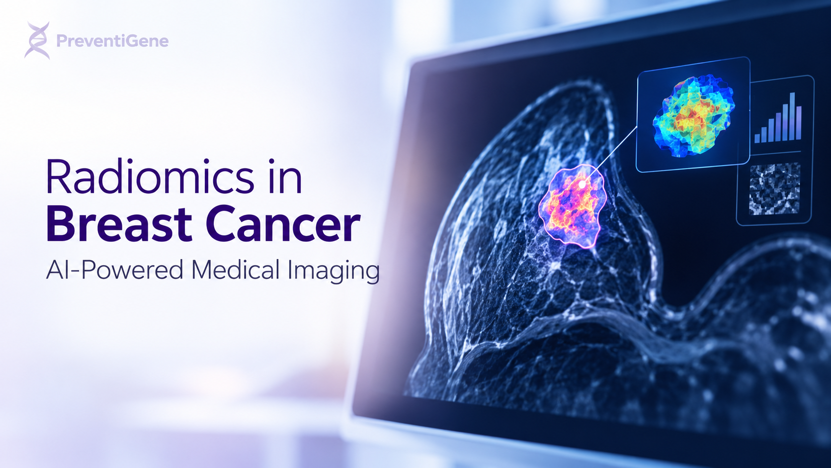
Radiomics is an advanced medical imaging approach that uses computational methods to extract large numbers of quantitative features from medical images. These features can describe tumor shape, size, texture, intensity patterns, and spatial heterogeneity—characteristics that may not be fully captured by conventional visual assessment alone.
By combining radiomics with artificial intelligence (AI), machine learning, and deep learning, researchers aim to identify imaging patterns associated with tumor biology, disease aggressiveness, treatment response, and clinical outcomes.
In breast cancer research, radiomics is being investigated as a promising component of precision oncology. Its objective is to complement established diagnostic methods and provide additional quantitative information that may help clinicians make more individualized decisions.
Importantly, radiomics is not a standalone diagnostic test. Its clinical value depends on rigorous validation, reproducibility, and integration with established clinical and pathological evidence.
How Does Radiomics Work?
A typical radiomics workflow involves several interconnected steps, from medical image acquisition to statistical analysis and model validation.
1. Medical Image Acquisition
Radiomics models can analyze images obtained through different imaging modalities, including:
Digital Mammography: Provides detailed breast images used in screening and diagnostic assessment.
Digital Breast Tomosynthesis (3D Mammography): Produces multiple image slices that help visualize breast structures with less overlap.
Breast Ultrasound: Provides information about lesion morphology and internal characteristics.
Breast Magnetic Resonance Imaging (MRI): Offers detailed information about breast tissue, tumor morphology, and contrast-enhancement patterns.
The suitability of each modality depends on the research question, imaging protocol, and clinical context. Different modalities provide different types of information and are not interchangeable.
2. Tumor Segmentation
The region of interest (ROI), such as a breast lesion, is identified within the medical image. This process is known as segmentation.
Segmentation may be performed manually by specialists, semi-automatically, or with AI-assisted tools. Accurate and reproducible segmentation is essential because errors at this stage can affect the quantitative features extracted later.
3. Quantitative Feature Extraction
Radiomics software converts images into measurable numerical features. Common feature categories include:
Shape Features: Describe tumor geometry, surface characteristics, and three-dimensional structure.
First-Order Statistics: Measure the distribution of image intensities within a selected region.
Texture Features: Quantify spatial patterns and relationships between image intensities.
Filtered Image Features: Reveal patterns at different spatial scales or frequency ranges.
Deep Learning Features: Learn complex image representations through neural networks rather than relying exclusively on predefined mathematical descriptors.
These measurements may capture aspects of tumor heterogeneity that are difficult to quantify consistently through visual inspection alone.
4. Artificial Intelligence and Predictive Modeling
Machine learning algorithms can analyze radiomic features alongside selected clinical variables to identify statistical associations with specific outcomes.
Depending on the study design, these models may investigate whether imaging patterns are associated with:
Benign versus malignant lesion classification.
Breast cancer molecular subtypes.
Histological grade and other pathological characteristics.
Response to neoadjuvant systemic therapy.
Recurrence risk and other prognostic outcomes.
Such associations do not automatically establish biological causation or guarantee accurate predictions for individual patients. Models must be evaluated using appropriate datasets and clinically relevant performance measures.
5. Model Validation and Clinical Interpretation
Before a radiomics model can be considered for clinical use, researchers need to evaluate its reliability and generalizability.
Good practice includes independent external validation, appropriate statistical analysis, calibration assessment, reproducible feature extraction, and evaluation across different scanners, institutions, and patient populations.
The Image Biomarker Standardisation Initiative (IBSI) provides standardized definitions and reference values for radiomic features. Reporting frameworks and quality assessment tools, including CLEAR and the Radiomics Quality Score, also help researchers assess study quality and transparency.
Applications of Radiomics in Breast Cancer
1. Breast Cancer Detection and Lesion Characterization
Radiomics research investigates whether quantitative imaging patterns can help distinguish malignant lesions from benign findings and improve the characterization of suspicious abnormalities.
AI-assisted analysis may identify patterns that complement conventional radiological assessment. However, radiomics should not replace established screening pathways, diagnostic imaging, or biopsy when clinically indicated.
2. Molecular Subtype Assessment
Breast cancer is a biologically diverse disease. Molecular and pathological characteristics, including estrogen receptor (ER), progesterone receptor (PR), HER2 status, and proliferation markers such as Ki-67, influence clinical management.
Researchers are investigating whether radiomic features correlate with these tumor characteristics. This field overlaps with radiogenomics, which explores relationships between medical imaging phenotypes and genomic or molecular information.
Although some studies report promising associations, imaging-based predictions cannot currently be assumed to replace tissue-based biomarker testing or other validated diagnostic procedures.
3. Treatment Response Prediction
Radiomics models can analyze baseline imaging and, in some studies, changes between imaging examinations to investigate associations with treatment response.
For example, breast MRI-derived features are being studied for their potential to predict response to neoadjuvant chemotherapy before surgery. Such research could eventually help identify patients who may benefit from different treatment strategies.
Nevertheless, the performance of these models varies, and prospective clinical validation is needed before they can reliably guide individual treatment decisions.
4. Prognostic Assessment
Tumor heterogeneity, morphology, and enhancement patterns may contain information associated with disease outcomes.
Researchers are studying whether radiomics can contribute to prognostic models that combine imaging findings with clinical variables, pathological results, and molecular biomarkers.
The aim is to develop more informative risk assessments, not to infer an individual's prognosis from imaging features alone.
5. Personalized Screening and Risk Assessment
AI-based breast imaging research also explores whether quantitative image analysis can contribute to more individualized screening strategies.
However, radiomics-based tumor characterization and population-level breast cancer risk assessment are different tasks. Imaging findings alone do not establish inherited cancer susceptibility, and radiomics should not be confused with germline genetic testing for variants in genes such as BRCA1 and BRCA2.
Personalized screening decisions should be based on established clinical guidance, individual risk factors, family history, and specialist assessment.
Radiomics, Radiogenomics, and Multi-Omics Integration
One of the most promising research directions is the integration of radiomics with genomic, transcriptomic, proteomic, and clinical data.
Each data type captures a different aspect of disease biology:
Radiomics: Quantifies spatial and structural patterns visible in medical images.
Genomics: Examines DNA variation and other genomic characteristics.
Transcriptomics: Investigates patterns of gene expression.
Proteomics: Studies proteins and their biological functions.
Clinical and Pathological Data: Provide patient-level and tissue-level information relevant to diagnosis and treatment.
Combining these sources may help researchers investigate how tumor appearance relates to its underlying molecular characteristics.
For example, breast MRI radiomic features may be evaluated alongside tumor gene-expression profiles and pathological biomarkers to study associations with tumor subtype or treatment response.
These approaches are part of a broader movement toward multimodal AI and precision medicine. Their success depends on reliable data integration, adequate sample sizes, independent validation, and careful assessment of potential bias.
The Role of AI in the Future of Intelligent Imaging
Traditional radiomics generally relies on predefined quantitative features, whereas deep learning can learn complex representations directly from medical images. Hybrid approaches combine these strategies with clinical and molecular information.
Current research directions include:
Deep Learning and Hybrid Models: Combining engineered radiomic features with learned image representations.
Multimodal AI: Integrating imaging, clinical records, pathology, and molecular data.
Explainable AI: Investigating which features contribute to model predictions and how reliable those explanations are.
Multi-Center Validation: Testing model performance across hospitals, scanners, and patient populations.
Standardized and Reproducible Workflows: Improving consistency in image processing, feature extraction, and model reporting.
Prospective Clinical Evaluation: Assessing whether radiomics provides meaningful benefits in real clinical settings.
The Radiomics Quality Score 2.0, published in Nature Reviews Clinical Oncology in 2025, reflects the growing emphasis on methodological rigor, fairness, explainability, quality control, and readiness for clinical translation.
These developments are important because a model that performs well in a retrospective research dataset may not perform equally well in everyday clinical practice.
Advantages and Limitations of Radiomics
Potential Advantages
Extracts quantitative information from existing medical images.
Enables systematic analysis of tumor heterogeneity.
May complement conventional imaging and pathological assessment.
Supports research into treatment response and prognostic biomarkers.
Creates opportunities to integrate imaging with genomic and clinical data.
May contribute to future personalized oncology workflows.
Current Limitations
Variability in imaging equipment, acquisition protocols, and reconstruction methods.
Differences in segmentation and feature extraction procedures.
Limited external validation for many published models.
Risk of overfitting, dataset bias, and poor generalizability.
Challenges in interpreting model predictions and establishing biological relevance.
Regulatory, privacy, workflow, and implementation requirements.
Radiomics should therefore be regarded as a developing research and clinical decision-support field rather than a universally established diagnostic technology.
How PreventiGene Fits into the Precision Medicine Pathway
At PreventiGene, our focus is on helping individuals better understand genetic health risks and navigate appropriate assessment and specialist care.
Radiomics and genetic testing provide different types of information, but they can be complementary within a broader precision medicine pathway.
Our role in this pathway includes:
1. Health Education and Initial Risk Awareness
PreventiGene provides educational information and online assessment tools designed to help individuals understand relevant health and hereditary risk factors.
Questionnaire-based assessments can support awareness and help identify when further professional evaluation may be appropriate. They do not establish a cancer diagnosis or independently confirm hereditary susceptibility.
2. Appropriate Genetic Assessment
When hereditary cancer risk is a concern, genetic assessment may be considered according to personal history, family history, and professional guidance.
Depending on the clinical context, testing may investigate relevant genes associated with inherited cancer susceptibility, including BRCA1 and BRCA2.
Genetic testing and radiomics answer different questions: genetic testing evaluates selected genetic variants, while radiomics analyzes quantitative patterns in medical images.
Neither should be interpreted in isolation when clinical decisions require a broader assessment.
3. Specialist Referral and Clinical Follow-Up
When an assessment indicates that further evaluation may be appropriate, PreventiGene can support referral to a relevant specialist.
Qualified healthcare professionals can determine whether additional imaging, genetic counseling, genetic testing, biopsy, or other investigations are appropriate.
Radiomics findings, when available from a clinically validated system, should be interpreted within this broader clinical context rather than treated as an independent diagnosis.
4. Connecting Emerging Technologies With Personalized Care
As AI-based imaging and multi-omics research advance, integrating reliable clinical, imaging, and genetic information may create new opportunities for more individualized healthcare.
PreventiGene's educational, assessment, testing-related, and referral pathway is intended to help individuals navigate appropriate next steps. Radiomics may be relevant to this evolving landscape, but it is not presented here as a diagnostic service currently provided by PreventiGene.
Our emphasis remains on scientifically grounded information, responsible use of health technologies, and appropriate professional guidance.
Frequently Asked Questions About Radiomics
Is radiomics used to diagnose breast cancer?
Radiomics is being investigated as a tool to support lesion characterization, risk stratification, and clinical decision-making. It is not a universal standalone diagnostic method and does not replace established imaging assessment, biopsy, or pathological examination when indicated.
Can radiomics replace breast biopsy?
No. Radiomics cannot currently be assumed to replace biopsy when tissue diagnosis is clinically necessary. Imaging-based predictions must be interpreted alongside established diagnostic evidence.
What is the difference between radiomics and radiogenomics?
Radiomics extracts quantitative features from medical images. Radiogenomics investigates relationships between imaging characteristics and genomic or molecular information. The two fields can overlap when researchers combine imaging features with molecular data.
Can radiomics predict breast cancer treatment response?
Some research models have shown associations between imaging features and treatment response. However, predictive performance varies, and independent validation and prospective clinical evaluation are needed before such models can be relied upon for individual treatment decisions.
Is radiomics the same as AI in medical imaging?
Not exactly. Radiomics is one approach to quantitative image analysis and may use machine learning to develop predictive models. AI in medical imaging is a broader field that also includes image detection, segmentation, classification, and deep learning methods that do not necessarily use conventional radiomic features.
Can radiomics identify inherited BRCA1 or BRCA2 variants?
No. Radiomics analyzes medical images and cannot independently confirm inherited BRCA1 or BRCA2 variants. Hereditary cancer assessment requires appropriate genetic evaluation, with professional guidance when indicated.
Conclusion
Radiomics and intelligent medical imaging represent important areas of research in precision oncology. By extracting quantitative features from breast imaging and combining them with artificial intelligence, clinical information, and molecular data, researchers are investigating new approaches to tumor characterization, treatment response assessment, and personalized care.
However, reproducibility, independent validation, interpretability, and demonstrated clinical benefit remain essential requirements for wider implementation.
For individuals concerned about hereditary cancer risk, radiomics should be understood as a complementary research field—not a substitute for appropriate screening, genetic assessment, or specialist care.
PreventiGene supports informed health decisions through education, risk-awareness tools, appropriate testing pathways, and specialist referral, helping individuals navigate the evolving landscape of personalized medicine.