Breast Ultrasound in Breast Cancer Detection and Diagnosis
Learn how contrast-enhanced mammography (CEM/CESM) helps detect and evaluate breast cancer, how the procedure works, its benefits, limitations, and role in modern breast imaging.
October 7, 2026
Breast Ultrasound in Breast Cancer Detection and Diagnosis
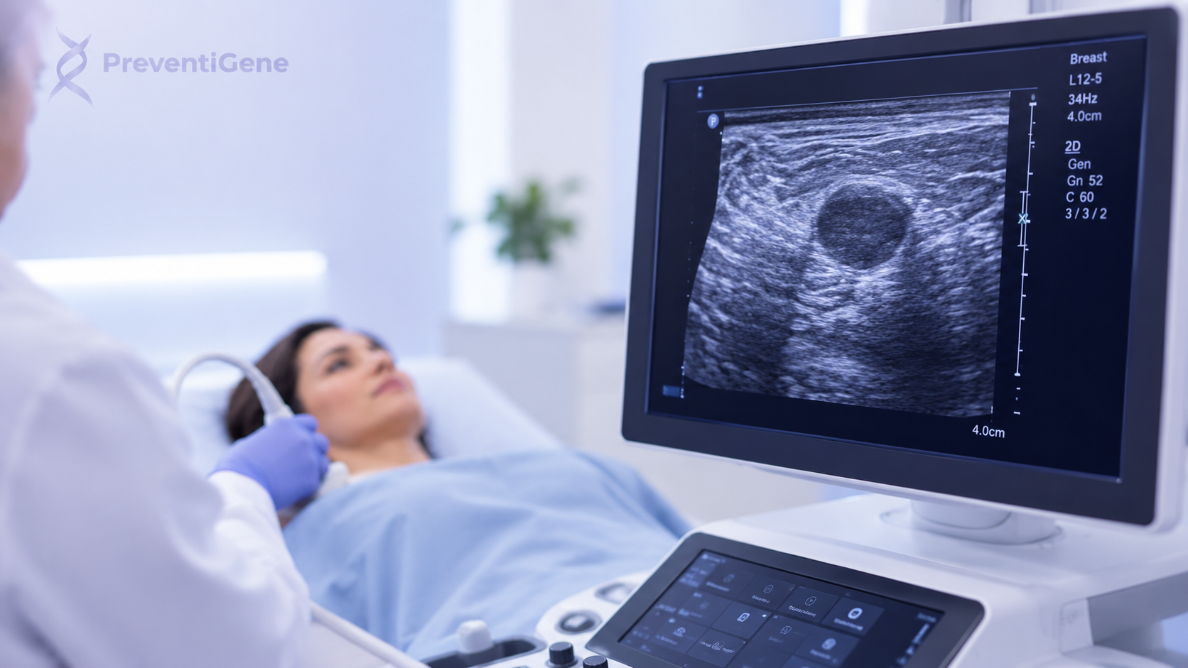
Breast ultrasound is an important imaging technique used to evaluate breast tissue and investigate abnormalities identified during a clinical examination or other breast imaging procedures. By using high-frequency sound waves, ultrasound produces real-time images of breast structures without exposing the patient to ionizing radiation.
It is particularly useful for characterizing breast masses, distinguishing fluid-filled cysts from solid lesions, and providing additional information when mammography findings require further assessment. However, breast ultrasound is not a replacement for mammography in routine breast cancer screening. Instead, the two techniques often complement each other as part of a broader breast imaging strategy.
At PreventiGene, we focus on helping individuals better understand their health and potential risk factors and navigate appropriate pathways for preventive and diagnostic care. When a breast abnormality or an elevated risk profile requires further evaluation, appropriate imaging and specialist assessment can become an important part of that pathway.
What Is a Breast Ultrasound?
A breast ultrasound, also known as breast sonography, uses a high-frequency transducer placed over the breast to create detailed images of the underlying tissues. Unlike mammography, ultrasound does not use X-rays or ionizing radiation.
During an examination, the radiologist can evaluate characteristics of a breast lesion, including its shape, margins, orientation, internal appearance, and relationship with surrounding tissues. These features can help determine whether a finding appears benign or suspicious and whether additional assessment may be necessary.
Breast ultrasound findings are commonly interpreted within standardized breast imaging frameworks such as the BI-RADS® (Breast Imaging Reporting and Data System), which helps radiologists communicate imaging findings and recommendations consistently.
When Is Breast Ultrasound Used?
Breast ultrasound may be recommended in several clinical situations, including:
Evaluation of a palpable breast lump
Assessment of focal breast pain or other localized symptoms
Further evaluation of an abnormality detected on mammography
Characterization of a breast mass as cystic or solid
Assessment of selected findings in dense breast tissue
Evaluation of specific breast abnormalities in younger patients
Assessment of abnormalities in the axillary region
Guidance for certain breast biopsy procedures
The appropriate imaging approach depends on factors such as age, symptoms, breast density, personal and family history, previous imaging findings, and overall breast cancer risk.
Breast Ultrasound and Dense Breasts
Breast density is an important consideration in breast imaging. Dense breast tissue contains a higher proportion of fibroglandular tissue, which can make certain abnormalities more difficult to visualize on mammography.
Ultrasound can provide additional information about some lesions that may be difficult to characterize on mammography. However, it should not automatically be considered a substitute for mammography. The decision to use supplemental ultrasound should take into account the patient's breast density, overall cancer risk, symptoms, and other clinical factors.
This is where individualized assessment becomes important. Rather than relying on a single imaging method, healthcare professionals can consider the patient's broader risk profile when determining whether additional breast imaging may be appropriate.
Can Breast Ultrasound Detect Breast Cancer?
Breast ultrasound can identify imaging features that may raise suspicion for breast cancer, but ultrasound alone cannot always determine whether a lesion is malignant.
Radiologists may assess characteristics such as:
Shape and orientation of the lesion
Margin characteristics
Echogenicity
Posterior acoustic features
Architectural distortion
Associated tissue changes
Other imaging features relevant to malignancy assessment
If a finding appears suspicious, additional diagnostic evaluation may be recommended. Depending on the situation, this can include diagnostic mammography, breast MRI, follow-up imaging, or tissue sampling.
The BI-RADS® assessment system provides a standardized framework for categorizing breast imaging findings and determining appropriate management.
Ultrasound-Guided Breast Biopsy
One of the important clinical applications of breast ultrasound is image-guided biopsy.
When a suspicious lesion can be clearly visualized using ultrasound, the imaging technique can guide a needle toward the target area during a biopsy. This allows tissue to be collected for pathological examination.
This distinction is important: imaging can identify and characterize a suspicious lesion, while pathology can determine whether cancer cells are present in the sampled tissue.
The combination of imaging, pathology, clinical information, and specialist assessment provides a more complete diagnostic picture.
Breast Ultrasound vs. Mammography
Breast ultrasound and mammography provide different types of information and are often complementary.
Mammography remains a fundamental breast imaging modality for screening and can be particularly valuable for detecting certain findings, including some microcalcifications that may not be visible on ultrasound.
Ultrasound, on the other hand, is especially useful for evaluating and characterizing breast masses and specific areas of concern. It can also provide valuable additional information in selected patients with dense breasts.
Depending on the clinical situation, breast cancer evaluation may therefore involve a combination of:
Mammography
Breast ultrasound
Digital breast tomosynthesis (3D mammography)
Breast MRI
Image-guided biopsy
Pathological examination
The appropriate combination depends on the individual patient's symptoms, risk factors, imaging findings, and clinical history.
Where PreventiGene Fits Into the Breast Cancer Care Pathway
Breast imaging is only one part of the broader breast cancer prevention and diagnostic pathway.
At PreventiGene, our role is to support individuals in understanding their health information, identifying relevant risk factors, and navigating appropriate next steps in preventive and diagnostic care.
For individuals concerned about breast cancer risk, this process may include reviewing relevant personal and family health information, considering appropriate risk assessment, and helping determine when consultation with a qualified healthcare professional or specialist may be appropriate.
When imaging is indicated, examinations such as mammography, breast ultrasound, breast MRI, or other diagnostic procedures should be performed and interpreted by qualified healthcare professionals. If an imaging result raises concern, appropriate specialist referral and, when necessary, biopsy and pathological evaluation are essential.
PreventiGene can serve as a bridge within this pathway by helping individuals move from health awareness and risk assessment toward appropriate medical evaluation and specialist care.
This approach is particularly relevant for individuals who may benefit from a more personalized understanding of their breast cancer risk rather than relying solely on population-based screening recommendations.
Integrating Imaging With Risk Assessment
Breast cancer risk is influenced by multiple factors, including age, family history, genetic predisposition, previous breast conditions, reproductive and hormonal factors, and other individual characteristics.
Imaging findings should therefore be interpreted within the broader clinical context.
For individuals with relevant personal or family histories, genetic risk assessment may also become an important component of preventive care. When appropriate, healthcare professionals may consider genetic counseling or genetic testing to investigate inherited susceptibility to certain cancers.
At PreventiGene, integrating health information and risk assessment with appropriate medical referral can help create a more individualized pathway. The goal is not to replace radiologists, oncologists, genetic counselors, pathologists, or other healthcare professionals, but to help individuals understand when additional evaluation may be warranted and navigate the next steps more effectively.
Why a Personalized Approach Matters
There is no single breast imaging strategy that is appropriate for every individual.
A person with a new breast lump may require targeted ultrasound. Another individual may benefit from mammography or 3D mammography as part of routine screening. Someone with a significantly elevated lifetime risk may require a different surveillance strategy, potentially involving breast MRI and specialist assessment.
For this reason, breast cancer detection should be approached as a risk-adapted and clinically guided process.
PreventiGene's role is to support this personalized approach by connecting health awareness, risk assessment, appropriate testing pathways, and professional medical care.
Conclusion
Breast ultrasound is a valuable component of modern breast imaging. It can help evaluate palpable abnormalities, characterize breast lesions, provide additional information in selected patients with dense breasts, and guide ultrasound-visible biopsies.
However, ultrasound should not be viewed as an independent replacement for mammography or other diagnostic methods. Accurate breast cancer evaluation may require a combination of imaging, clinical assessment, pathology, and—when appropriate—genetic and risk assessment.
At PreventiGene, our focus is on helping individuals understand their potential health risks and navigate the appropriate pathway toward preventive care, diagnostic evaluation, and specialist referral.
By combining informed risk assessment with appropriate medical imaging and professional evaluation, individuals can take a more proactive and personalized approach to breast health and breast cancer prevention.