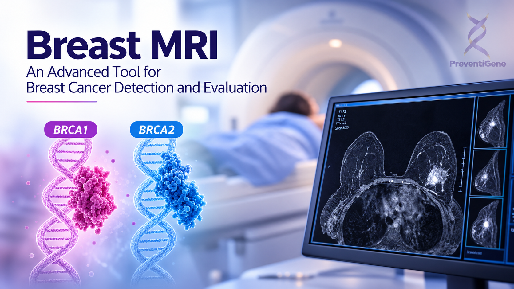
Learn how breast MRI supports breast cancer detection, high-risk screening, diagnosis, and treatment planning, and how PreventiGene supports personalized cancer risk assessment.
October 7, 2026
Breast MRI: An Advanced Tool for Breast Cancer Detection and Evaluation
Breast cancer detection increasingly relies on a combination of advanced imaging technologies, clinical assessment, and individualized risk evaluation. Among these technologies, breast MRI is one of the most sensitive imaging methods available for identifying and evaluating breast abnormalities.
Breast MRI uses a powerful magnetic field, radiofrequency waves, and computer processing to create detailed images of breast tissue. Unlike mammography, MRI does not use ionizing radiation. When performed with contrast enhancement, it can highlight areas with increased vascular activity and help radiologists identify suspicious lesions that may not be clearly visible on conventional breast imaging.
However, breast MRI is not intended to replace mammography, ultrasound, or biopsy. Instead, it is used as a complementary imaging technique for specific patients and clinical situations.
What Is Breast MRI?
Breast magnetic resonance imaging (MRI) is a non-invasive imaging technique that produces detailed, cross-sectional images of the breasts.
For most breast cancer evaluations, a gadolinium-based contrast agent is administered intravenously during the examination. The contrast material helps demonstrate differences in blood flow and tissue enhancement, allowing suspicious areas to be assessed more clearly.
During the examination, the patient lies face down on a specialized MRI table while the breasts are positioned within a dedicated breast coil. Multiple imaging sequences are obtained before and after contrast administration.
Because MRI does not use X-rays, it provides detailed breast imaging without exposure to ionizing radiation.
When Is Breast MRI Used?
Breast MRI is not routinely recommended as the first-line screening test for every woman. Its use depends on factors such as breast cancer risk, symptoms, previous imaging findings, breast density, personal history, and genetic or familial risk.
1. Screening Women at High Risk of Breast Cancer
One of the most important applications of breast MRI is screening women who have a significantly increased risk of developing breast cancer.
Depending on individual risk assessment and clinical guidelines, MRI may be recommended in addition to mammography for women with factors such as:
A lifetime breast cancer risk of approximately 20% or higher
Certain inherited genetic variants associated with breast cancer
A strong family history of breast or ovarian cancer
A personal history of breast cancer
Previous radiation therapy to the chest at a young age
For these individuals, combining different imaging modalities can increase the likelihood of detecting clinically significant abnormalities.
2. Evaluating Suspicious Findings
Mammography or ultrasound may sometimes reveal an abnormality that cannot be adequately characterized using conventional imaging.
In selected cases, breast MRI can provide additional information about the lesion, including its location, size, morphology, and enhancement pattern. This additional information can help physicians determine whether further diagnostic evaluation or biopsy should be considered.
MRI findings must always be interpreted in combination with the patient's clinical history and other imaging results.
3. Determining the Extent of Known Breast Cancer
After breast cancer has been diagnosed, MRI may be used to assess the extent of disease within the breast.
It can help identify:
The size and location of the primary tumor
Additional lesions within the same breast
Possible abnormalities in the opposite breast
The relationship between a lesion and surrounding breast tissue
This information can contribute to treatment planning and surgical decision-making.
4. Evaluating the Breast After Treatment
Breast MRI can also be useful in selected patients who have previously undergone breast cancer treatment.
After surgery or radiation therapy, scar tissue and recurrent cancer can sometimes produce similar findings on conventional imaging. MRI may provide additional information when mammography, ultrasound, or clinical examination cannot adequately explain a new or changing abnormality.
Breast MRI vs. Mammography and Ultrasound
No single imaging technique can answer every clinical question in breast cancer detection.
Mammography uses low-dose X-rays and remains one of the most important tools for breast cancer screening. It is particularly valuable for detecting masses and suspicious calcifications.
Breast ultrasound uses sound waves and is commonly used to evaluate palpable abnormalities and further characterize findings detected on mammography. It can help differentiate cystic from solid lesions.
Breast MRI provides highly detailed images and has particularly high sensitivity for detecting breast cancer. It is especially valuable for selected high-risk patients and for evaluating the extent of known disease or unresolved findings.
These technologies should therefore be considered complementary rather than competing tests.
How Accurate Is Breast MRI for Breast Cancer Detection?
Breast MRI is known for its high sensitivity in detecting breast cancer. It can reveal some cancers that are not clearly visible on mammography or ultrasound.
However, high sensitivity does not mean that every MRI finding represents cancer.
Benign lesions and normal variations in breast tissue can sometimes enhance after contrast administration and appear suspicious. This may lead to additional imaging, follow-up examinations, or biopsy.
For this reason, MRI results should be interpreted by experienced breast imaging specialists and considered together with the patient's clinical and imaging history.
What Happens If Breast MRI Shows a Suspicious Lesion?
An abnormal MRI finding does not automatically mean that breast cancer is present.
Depending on the characteristics of the lesion, the next step may include:
Additional mammographic or ultrasound evaluation
Short-term imaging follow-up in selected cases
MRI-guided biopsy when the abnormality cannot be adequately evaluated using another imaging modality
A biopsy provides tissue for pathological examination and is required when a definitive tissue diagnosis is necessary.
This illustrates an important principle in modern breast cancer detection: imaging is part of a diagnostic pathway rather than a standalone diagnosis.
The Role of Risk Assessment in Breast MRI
The decision to perform breast MRI should ideally be based on more than a single symptom or imaging finding.
A comprehensive assessment may consider:
Personal and family history of cancer
Age and other clinical characteristics
Previous breast imaging
Breast density
Known or suspected hereditary cancer risk
Previous cancer treatment
Current symptoms and clinical findings
At PreventiGene, we focus on supporting a more individualized approach to cancer prevention and early detection. By integrating health information, risk assessment, and appropriate referral pathways, our goal is to help individuals and healthcare professionals make better-informed decisions about when additional evaluation or specialist consultation may be appropriate.
PreventiGene does not replace radiologists, oncologists, surgeons, or other healthcare professionals. Instead, our role is to support the pathway between risk awareness, appropriate assessment, diagnostic services, and specialist care.
How PreventiGene Supports the Breast Cancer Detection Pathway
Modern cancer prevention is not simply about performing more tests. The goal is to identify the right assessment for the right individual at the right time.
PreventiGene can contribute to this process by helping individuals understand their potential risk factors, providing structured health and lifestyle assessments, supporting access to appropriate genetic and health-related testing when indicated, and facilitating referral to qualified healthcare professionals.
When breast imaging is required, the information obtained through risk assessment and clinical evaluation can help inform discussions with healthcare providers about whether conventional imaging or advanced modalities such as breast MRI may be appropriate.
For individuals with concerning findings, the pathway may continue from imaging to specialist assessment and, when clinically indicated, tissue diagnosis.
This integrated approach helps place breast MRI within the broader framework of personalized breast cancer prevention and early detection, rather than treating MRI as an isolated diagnostic test.
Benefits of Breast MRI
Breast MRI offers several important advantages:
High sensitivity for detecting breast cancer
No exposure to ionizing radiation
Detailed visualization of breast tissue
Useful supplemental screening for women at elevated risk
Assessment of the extent of known breast cancer
Evaluation of selected abnormalities that remain unclear after conventional imaging
Ability to guide biopsy of lesions visible only on MRI
Limitations of Breast MRI
Despite its advantages, breast MRI also has important limitations.
MRI can produce false-positive findings, meaning that an area may appear suspicious even though it is ultimately benign. This can result in additional imaging or unnecessary biopsies.
Contrast-enhanced MRI usually requires intravenous administration of a gadolinium-based contrast agent. The patient's medical history, including relevant kidney conditions, pregnancy status, allergies, and implanted medical devices, should be discussed with the healthcare provider before the examination.
MRI can also be more expensive and less widely available than conventional breast imaging. In addition, the patient must remain relatively still during the examination, which can be challenging for some individuals.
Breast MRI as Part of Personalized Breast Cancer Detection
Breast cancer detection is increasingly moving toward personalized strategies that combine clinical information, risk assessment, imaging, genetics, and specialist evaluation.
Breast MRI has an important role within this approach, particularly for women at elevated risk or for situations where conventional imaging does not provide sufficient information.
However, choosing the appropriate imaging method should always be based on the individual's risk profile, symptoms, previous findings, and clinical recommendations.
At PreventiGene, our approach is centered on connecting risk awareness and personalized assessment with appropriate healthcare pathways. By helping individuals better understand their risk and supporting access to relevant testing and specialist referral, we aim to contribute to earlier awareness and more informed decisions throughout the breast cancer detection journey.
Key Takeaway
Breast MRI is a highly sensitive, radiation-free imaging technique that can provide valuable information for high-risk breast cancer screening and the evaluation of suspicious or previously diagnosed breast cancer. It is most effective when used as part of a broader, personalized diagnostic pathway alongside mammography, ultrasound, clinical assessment, and—when necessary—biopsy.