Learn how breast biopsy confirms breast cancer, the main biopsy types, pathology testing, and how PreventiGene supports patients through the diagnostic pathway.
October 8, 2026
Breast Biopsy in Breast Cancer Diagnosis
A breast biopsy is one of the most important steps in the diagnosis of breast cancer. Modern breast imaging techniques, including mammography, 3D mammography, ultrasound, and breast MRI, can identify and characterize suspicious abnormalities. However, imaging alone cannot definitively determine whether an abnormality is cancerous.
When imaging or clinical examination identifies a suspicious area, a biopsy may be recommended to obtain a sample of breast tissue or cells. The sample is then examined by a pathologist to determine whether cancer is present and, when applicable, to characterize the biological features of the tumor.
For this reason, breast biopsy plays a central role in moving from suspicion to a tissue-based diagnosis.
What Is a Breast Biopsy?
A breast biopsy is a procedure in which cells, tissue, or part of a suspicious breast lesion is removed for laboratory examination.
A biopsy may be recommended after a mammogram, ultrasound, MRI, or clinical examination detects a finding that requires further investigation. Importantly, being referred for a biopsy does not mean that a person has breast cancer. Many breast abnormalities evaluated by biopsy are benign.
The purpose of the biopsy is to obtain direct biological evidence that can help distinguish between benign, atypical, in situ, and invasive lesions.
Why Is Biopsy Important in Breast Cancer Diagnosis?
Breast imaging provides essential information about the location, size, shape, and appearance of an abnormality. It can help radiologists determine whether a finding appears benign or suspicious and whether tissue sampling should be considered.
However, imaging cannot replace pathological examination.
During a biopsy, tissue is obtained from the area of concern and sent to a pathology laboratory. A pathologist examines the specimen under a microscope and prepares a pathology report. If cancer is identified, additional testing may be performed to characterize the tumor and support treatment planning.
The diagnostic pathway can therefore be summarized as:
Breast imaging → Suspicious finding → Image-guided biopsy → Pathological examination → Diagnosis and tumor characterization
When Is a Breast Biopsy Recommended?
A healthcare professional may recommend a biopsy when an abnormality cannot be confidently classified through imaging or clinical examination alone.
Examples include:
A suspicious breast mass
Microcalcifications detected on mammography
Architectural distortion
A suspicious lesion identified by ultrasound
An abnormal area detected on breast MRI
A palpable abnormality with concerning characteristics
A finding that requires tissue confirmation
The decision to perform a biopsy depends on the individual clinical situation and the characteristics of the abnormality.
Types of Breast Biopsy
Different biopsy techniques are available, and the most appropriate method depends on the location and characteristics of the lesion and which imaging modality best visualizes it.
Core Needle Biopsy
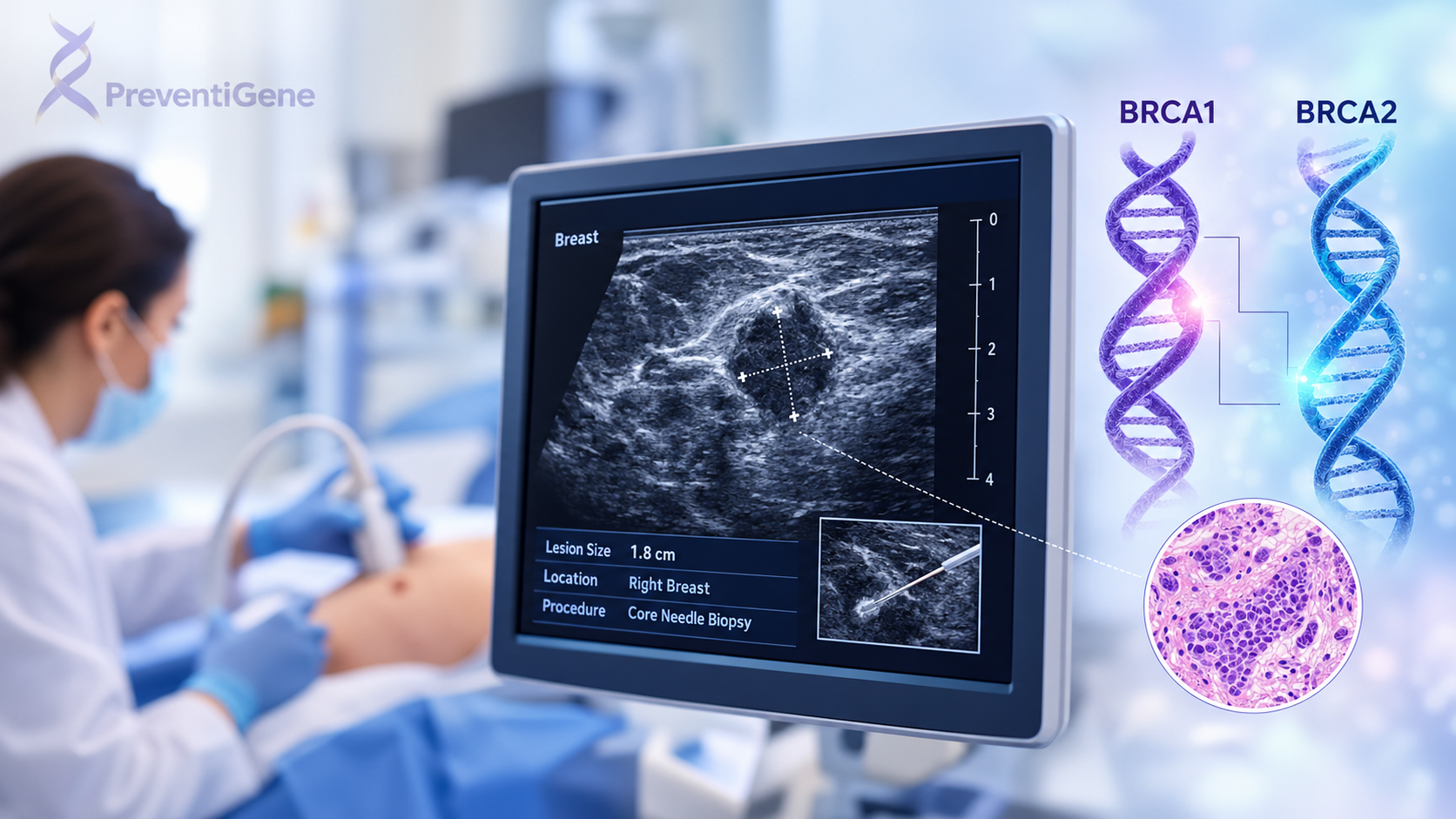
Core needle biopsy (CNB) is one of the most commonly used methods when breast cancer is suspected.
A hollow needle is used to remove several small cylindrical samples of tissue from the suspicious area. Unlike fine-needle aspiration, core biopsy provides tissue architecture, which can allow more detailed pathological evaluation and additional testing when needed.
Core needle biopsy can be guided by:
Ultrasound
Mammography or tomosynthesis
MRI
The imaging modality selected depends on where and how the suspicious abnormality can best be visualized.
Ultrasound-Guided Breast Biopsy
When a suspicious lesion can be clearly seen on ultrasound, ultrasound may be used to guide the biopsy needle to the precise location.
This approach allows the physician to visualize the target in real time while collecting tissue samples.
Stereotactic Breast Biopsy
A stereotactic breast biopsy uses mammographic images from different angles to precisely locate an abnormality.
It is particularly useful for certain findings, including suspicious microcalcifications or abnormalities that are visible on mammography but are not adequately visualized by ultrasound.
This is one example of how breast imaging and biopsy work together: imaging identifies and localizes the target, while biopsy provides tissue for pathological confirmation.
MRI-Guided Breast Biopsy
Some abnormalities are visible on breast MRI but cannot be adequately targeted using mammography or ultrasound.
In these situations, MRI-guided biopsy may be used. MRI helps the physician accurately localize the suspicious area and guide the biopsy device to the target.
Fine-Needle Aspiration
Fine-needle aspiration (FNA) uses a very thin needle to remove cells or fluid from a suspicious area.
FNA can be useful in selected situations, including some cystic lesions. However, when a solid breast lesion requires more extensive tissue evaluation, core needle biopsy is often preferred because it provides a larger tissue sample.
Surgical Biopsy
In selected cases, a surgical biopsy may be performed to remove part or all of an abnormal area.
Surgical biopsy may be:
Incisional biopsy: removal of part of the abnormal area
Excisional biopsy: removal of the entire abnormal area
Most breast biopsies are performed using needle-based techniques, while surgical biopsy is reserved for selected clinical situations.
What Happens During a Breast Biopsy?
The exact process depends on the biopsy technique.
For many image-guided needle biopsies, the patient receives local anesthesia to numb the area. The physician then uses the appropriate imaging modality to locate the suspicious lesion and guide the biopsy needle.
Several tissue samples may be collected and sent to the pathology laboratory.
A small marker clip may also be placed at the biopsy site. This marker can help healthcare professionals identify the location later during follow-up imaging or additional procedures.
Needle biopsies are generally performed as outpatient procedures, although the exact setting and recovery depend on the type of biopsy performed.
What Happens to the Biopsy Sample?
The biopsy specimen is sent to a pathology laboratory, where it undergoes tissue processing and microscopic examination.
The pathologist evaluates the cellular and structural characteristics of the tissue and determines whether the findings are benign or malignant.
If cancer is identified, the pathology report may provide important information such as:
Type of breast cancer
Whether the tumor is invasive or non-invasive
Tumor grade
Tissue characteristics
Hormone receptor status
HER2 status
Other clinically relevant pathological findings
These findings can provide important information for determining the most appropriate next steps in patient management.
Breast Biopsy and Biomarker Testing
The role of biopsy does not necessarily end with determining whether cancer is present.
When breast cancer is diagnosed, the biopsy tissue may be used for biomarker testing. Depending on the clinical situation, testing may include estrogen receptor (ER), progesterone receptor (PR), and HER2.
These biomarkers provide information about the biological characteristics of the tumor and can influence treatment decisions.
This is one reason obtaining an adequate tissue sample is important: the biopsy can provide both diagnostic and biological information.
How Breast Imaging and Biopsy Work Together
Breast cancer diagnosis is not based on a single test. Different diagnostic tools answer different questions.
Mammography and 3D mammography can identify suspicious structural changes and calcifications.
Breast ultrasound can help characterize masses and can be particularly useful for evaluating palpable or mammographically detected abnormalities.
Breast MRI can provide detailed information about breast tissue and can identify certain lesions that may not be adequately characterized by other imaging techniques.
When tissue confirmation is necessary, biopsy provides the sample required for pathological diagnosis.
Therefore, these technologies should not be viewed as competing diagnostic methods. They are complementary components of a broader diagnostic pathway.
The Role of PreventiGene in the Breast Cancer Diagnostic Pathway
At PreventiGene, our role is not to replace the radiologist, breast surgeon, pathologist, or treating oncologist. Instead, we aim to support individuals throughout the broader pathway of breast cancer awareness, risk assessment, diagnostic evaluation, and informed decision-making.
When a breast abnormality is identified, understanding what happens next can be difficult and overwhelming. PreventiGene can help patients better understand the purpose of recommended assessments and how different stages of the diagnostic pathway fit together.
Our role may include:
Supporting Awareness and Early Evaluation
PreventiGene provides evidence-based information to help individuals understand breast cancer risk factors, screening, symptoms, and available diagnostic approaches.
Recognizing when further medical evaluation may be appropriate is an important part of responsible health management.
Helping Navigate the Diagnostic Pathway
Breast cancer diagnosis can involve several professionals and technologies, from breast imaging to biopsy and pathology.
PreventiGene can help patients understand the relationship between these steps and identify when consultation with an appropriate healthcare professional or specialist may be necessary.
Connecting Diagnostic Information
A biopsy result should not be interpreted in isolation.
Pathology findings are considered together with imaging results, clinical history, physical examination, and other relevant information. PreventiGene's approach focuses on helping patients understand how these different sources of information contribute to the broader clinical picture.
Supporting Informed Next Steps
If a biopsy confirms breast cancer, additional evaluation may be required to characterize the tumor and determine its extent and biological features.
PreventiGene can support patients in understanding the questions they may need to discuss with their physician or specialist and in navigating the next stages of their healthcare journey.
From Detection to Diagnosis: A Multidisciplinary Process
A breast abnormality may be detected during screening or because of symptoms. From that point, several complementary steps may be required.
A simplified pathway is:
Risk assessment and awareness
↓
Breast imaging
↓
Identification of a suspicious finding
↓
Specialist evaluation
↓
Image-guided or surgical biopsy when indicated
↓
Pathological examination
↓
Biomarker and additional testing when appropriate
↓
Clinical assessment and treatment planning
No single test provides every piece of information required for breast cancer management. Instead, diagnosis and treatment planning depend on integrating clinical, imaging, pathological, and biological information.
Final Thoughts
A breast biopsy is a critical diagnostic procedure when breast imaging or clinical evaluation identifies a suspicious abnormality. While mammography, 3D mammography, ultrasound, and MRI can detect and characterize abnormalities, biopsy provides the tissue required for pathological confirmation.
Core needle biopsy is commonly used, while ultrasound-guided, stereotactic, MRI-guided, fine-needle, and surgical techniques may be selected according to the individual case.
The information obtained from biopsy and subsequent pathology and biomarker testing can help establish a more precise diagnosis and provide important information for clinical decision-making.
At PreventiGene, we believe that understanding the diagnostic pathway is an important part of informed healthcare. Our goal is to help individuals navigate that pathway with reliable information, appropriate professional guidance, and a clearer understanding of the steps between detection, diagnosis, and personalized care.