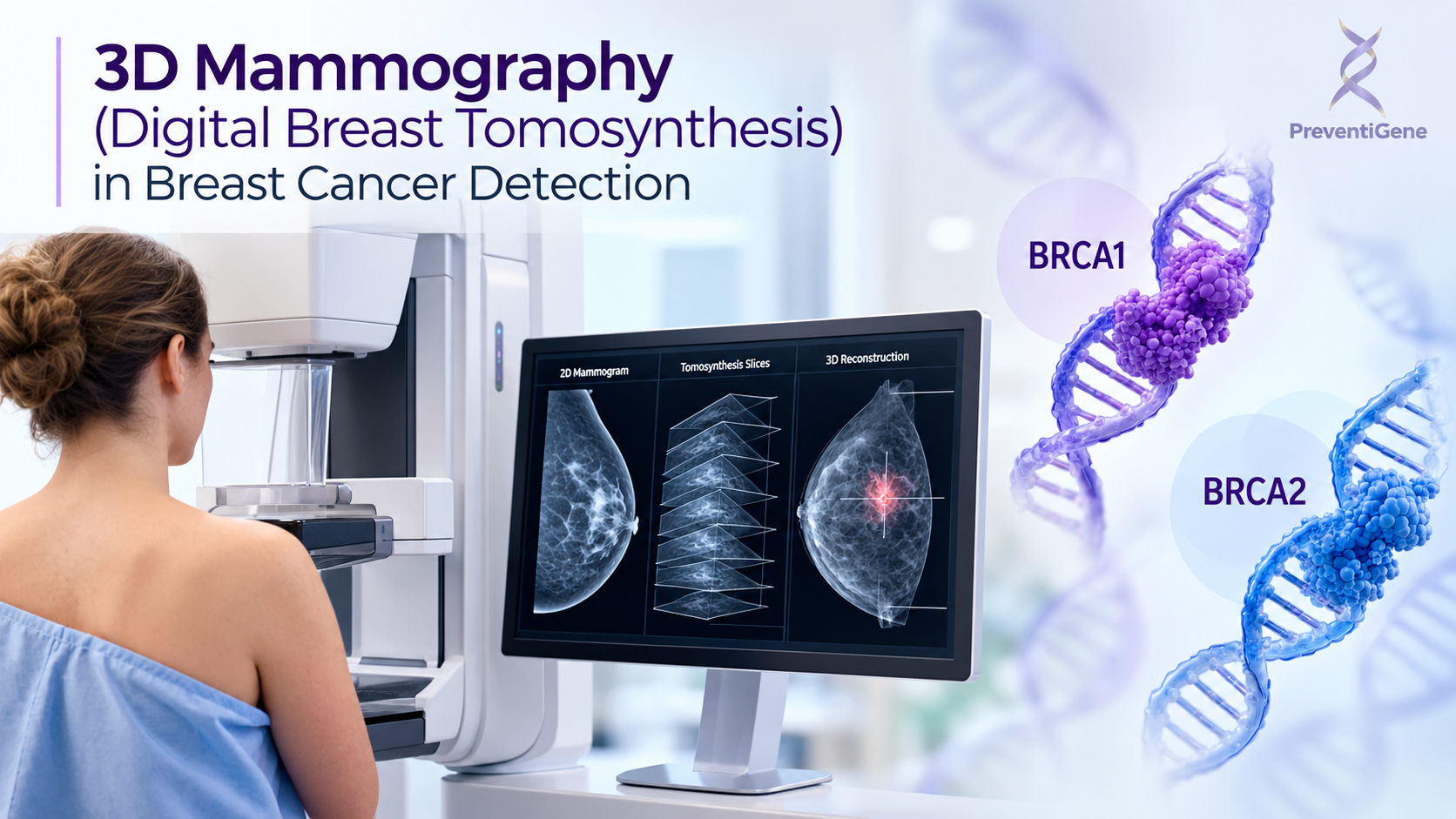
Discover how 3D mammography (digital breast tomosynthesis) enhances breast imaging and supports breast cancer detection, particularly in dense breasts, as part of a personalized breast health and screening pathway.
October 6, 2026
3D Mammography in Breast Cancer Detection
Early detection is an important component of breast cancer care. Modern breast imaging technologies have expanded beyond conventional mammography to provide more detailed visualization of breast tissue and support the identification of suspicious abnormalities.
One of these technologies is 3D mammography, medically known as digital breast tomosynthesis (DBT). By acquiring multiple low-dose X-ray images from different angles and reconstructing them into thin slices, 3D mammography allows breast tissue to be evaluated layer by layer rather than as a single two-dimensional image.
For appropriate patients, this technology can provide additional information during breast cancer screening and diagnostic evaluation, particularly when overlapping breast tissue makes abnormalities more difficult to visualize.
What Is 3D Mammography?
Traditional digital mammography produces two-dimensional images of the breast. Although these images are highly valuable for breast cancer screening, normal breast structures can overlap on a 2D image and potentially obscure an abnormality.
Digital breast tomosynthesis (DBT) addresses this limitation by taking multiple X-ray projections while the X-ray tube moves through an arc around the compressed breast. A computer then reconstructs these projections into a series of thin images.
The radiologist can review these slices individually, allowing breast structures to be examined at different depths. This can make certain masses, architectural distortions, and other abnormalities easier to distinguish from surrounding tissue.
How Does 3D Mammography Work?
The procedure is similar to a conventional mammogram. The breast is positioned and compressed between the imaging plates, while the X-ray system acquires multiple low-dose images from different angles.
These images are then reconstructed into a three-dimensional representation of the breast.
The resulting images do not represent a literal three-dimensional photograph. Instead, they provide a series of thin reconstructed sections that allow the radiologist to examine the breast tissue layer by layer.
This reduction in tissue overlap is one of the key reasons 3D mammography can provide additional information compared with conventional 2D mammography.
3D Mammography vs. 2D Mammography
Both technologies use X-rays to examine breast tissue, but the way information is acquired differs.
A conventional mammogram produces a small number of two-dimensional projections. Digital breast tomosynthesis, on the other hand, acquires multiple projections and reconstructs them into thin slices.
This can help reduce the masking effect caused by overlapping tissue. Clinical evidence has shown that DBT can increase cancer detection in some screening populations and reduce recall rates compared with conventional 2D mammography alone.
However, 3D mammography does not eliminate the limitations of breast imaging. Some cancers may remain difficult to detect, and an abnormal result does not necessarily mean that cancer is present.
Why Can 3D Mammography Be Helpful for Dense Breasts?
Breast density is an important consideration in breast cancer screening.
Dense breasts contain a greater proportion of fibroglandular tissue. On a mammogram, both dense tissue and many breast abnormalities can appear white. This can make some lesions more difficult to distinguish from the surrounding tissue.
Because DBT generates multiple thin slices, it can reduce the visual overlap of breast structures and may improve the visibility of certain abnormalities.
However, breast density alone does not determine the most appropriate screening strategy. Depending on an individual's age, personal and family history, genetic risk, symptoms, and other factors, healthcare professionals may consider additional imaging modalities such as ultrasound or breast MRI.
Can 3D Mammography Diagnose Breast Cancer?
3D mammography is primarily an imaging and detection tool. It can identify suspicious findings, but it cannot by itself confirm that a suspicious lesion is cancer.
When an abnormality is detected, further evaluation may include:
Additional mammographic views
Breast ultrasound
Breast MRI in selected situations
Image-guided biopsy when clinically indicated
Pathological examination of tissue obtained through biopsy
A definitive diagnosis of breast cancer generally requires pathological assessment when tissue sampling is indicated.
This distinction is important: an imaging finding is not the same as a cancer diagnosis.
What Are the Advantages of 3D Mammography?
Depending on the clinical and screening context, digital breast tomosynthesis may offer several advantages:
More detailed visualization of breast structures
Reduced impact of overlapping tissue
Improved detection of some breast cancers compared with 2D mammography alone
Fewer false-positive recalls in many screening settings
Potential advantages for some women with dense breast tissue
At the same time, DBT has limitations. It still involves exposure to a small amount of ionizing radiation, does not detect every breast cancer, and may lead to additional imaging or biopsy when an abnormality requires further investigation.
Therefore, the choice of imaging technique should be individualized rather than based on the assumption that one modality is universally appropriate for everyone.
Where Does 3D Mammography Fit in the Breast Cancer Detection Pathway?
Breast cancer detection is not based on a single test. Depending on an individual's risk profile and clinical circumstances, the pathway may involve several complementary stages:
Risk assessment → Screening → Breast imaging → Further diagnostic evaluation → Specialist assessment → Biopsy and pathology when indicated
3D mammography can play an important role within the breast imaging stage of this pathway.
Its findings can help determine whether additional investigation is necessary, but interpretation should always be integrated with the patient's clinical history and other available information.
The Role of PreventiGene in the Breast Health Journey
At PreventiGene, our role is not to replace breast imaging or specialist diagnosis. Instead, we aim to support individuals in navigating a more informed and personalized approach to health and disease prevention.
Breast health assessment can begin before an imaging abnormality is identified. Understanding personal and family history, evaluating relevant risk factors, and considering genetic risk when appropriate can help inform discussions about screening and preventive strategies.
When further assessment is indicated, PreventiGene can help individuals understand available options and navigate the next steps, including appropriate referral to physicians and specialists.
This approach places 3D mammography and other diagnostic technologies within a broader, personalized healthcare pathway, rather than treating any single test as a standalone answer.
3D Mammography as Part of Modern Breast Cancer Detection
Digital breast tomosynthesis represents an important development in breast imaging. By providing multiple reconstructed views through the breast, it can reduce the effect of overlapping tissue and help radiologists identify certain abnormalities that may be less conspicuous on conventional mammography.
Nevertheless, breast cancer detection requires a broader clinical perspective. Mammography, ultrasound, MRI, genetic risk assessment, specialist evaluation, and pathology each have different roles and should be used according to the individual's circumstances.
At PreventiGene, we focus on connecting risk awareness, personalized assessment, appropriate screening, and specialist care to help individuals make informed decisions about their breast health.
If you have concerns about your breast cancer risk, family history, breast density, or the appropriate screening strategy for you, discuss your individual situation with a qualified healthcare professional.